![Search[Google]](../../common/image/hmenu_search.png)

![CMIS[RIKEN Center for Molecular Imaging Science]RIKEN Kobe Institute Center for Molecular Imaging Science](../../common/image/hlogo_txt.png)


When we move our arms or legs, a molecule called dopamine functions to mediate the exchange of necessary signals among neurons in the brain. Degeneration of dopamine-producing neurons caused by some reasons will inhibit smooth regulation of body movement, and cause motor dysfunction. One of the typical diseases with this symptom is Parkinson's disease, which is an intractable disease without a curative treatment because lost nerves cannot be regenerated. Recent advancement in stem cell study, however, has led to the expectation that regenerative therapy with transplantation will be realized to replace lost dopaminergic neurons. Induced pluripotent stem cells (iPSCs) developed by Dr. Shinya Yamanaka of Kyoto University, et al., in particular, allowed transplantation of autologous cells to be carried out without rejection, and are currently under study for clinical application in many countries.
In a transplantation therapy using iPSCs, accurate monitoring on the conditions of transplanted cells or the brain surrounding them is required to check whether transplanted cells function as expected, whether they do not act as cancerous cells, or whether there is any rejection. This monitoring should be continued for a long time beginning just after the transplantation, which calls for a simple and patient-friendly method. The accurate live imaging of transplanted cells with a non-invasive diagnostic imaging technique such as PET or MRI will be significant progress toward the clinical application of transplantation therapy.
Our study team explored the culture conditions that allowed human iPSCs to efficiently turn into dopaminergic neurons. Then, the dopaminergic neurons generated under these conditions were transplanted into a cynomolgus monkey in which the symptoms of Parkinson's disease were artificially induced by administering a dopaminergic neurotoxin. First of all, the positions of transplanted cells were identified on histological MRI images to examine the post-transplant graft survival. Then, dopamine precursor molecules labeled with radioisotope were administered, and tested by PET. The results showed an aggressive intake of the molecules by the transplanted cells. An experiment using another labeling compound confirmed the absence of abnormal proliferation of the transplanted cells. These results demonstrate that the transplanted cells deriving from human iPSCs function as normal neurons with dopamine-producing capacity in the monkey brain. These findings were also verified in histopathological studies.
The results of this study demonstrated that cell transplantation therapy with iPSCs may be an effective treatment with a good safety, and that molecular imaging techniques including MRI and PET are very useful evaluation tools to verify the results of transplantation therapy in a non-invasive manner. We are going to proceed with the study in order to refine the techniques so that a more specific live imaging of transplanted cells will be available, and to use these techniques also in human application.
* This study was a joint program between a study group in Center for iPS Cell Research and Application, Kyoto University (researcher Tetsuhiro Kikuchi, associate professor Jun Takahashi) and a team in Functional Probe Research Laboratory, RIKEN Center for Molecular Imaging Science (team leader Hirotaka Onoe, deputy team leader Takuya Hayashi, researcher Toshiyuki Kawasaki). See here for more details of the study.
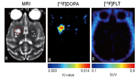 |
| MRI image (left) and PET images (center, right) showing the brain of Parkinson's disease model monkey six months after transplantation of iPSC-derived dopaminergic neurons. In the region in which the engraftment of transplanted cells was detected by MRI image (red circle), dopamine-producing capacity (center) and absence of tumorigenesis (right) were shown on PET images. [18F]DOPA is the dopamine precursor molecule DOPA labeled with fluorine radioisotope 18F, and [18F]FLT is a molecule in which DNA-comprising thymidine is labeled with 18F. Both are PET probes with which to detect dopamine-producing cells and dividing cell (tumor cells), respectively. The area with high level probe accumulation is highlighted red on PET images. |
