New research uncovers the neural mechanism underlying drug cravings
Cues such as the sight of drugs can induce cravings and lead to drug-seeking behaviors and drug use. But cravings are also influenced by other factors, such as drug availability and self-control. To investigate the neural mechanisms involved in cue-induced cravings, research group collaborating with Professor Alain Dagher from the Montreal Neurological Institute of McGill University in Canada studied the brain activity of a group of 10 smokers, following exposure to cigarette cues under two different conditions of cigarette availability. In one experiment cigarettes were available immediately and in the other they were not. We combined a technique called transcranial magnetic stimulation (TMS) with functional magnetic resonance imaging (fMRI).
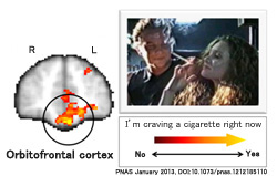
The results demonstrate that in smokers the orbitofrontal cortex (OFC) tracks the level of craving while the dorsolateral prefrontal cortex (DPFC) is responsible for integrating drug cues and drug availability. Moreover, the DPFC has the ability to suppress activity in the OFC when the cigarette is unavailable. When the DPFC was inactivated using TMS, both craving and craving-related signals in the OFC became independent of drug availability.
It would be considered that the DLPFC incorporates drug cues and knowledge on drug availability to modulate the value signals it transmits to the OFC, where this information is transformed into drug-seeking action.
These findings will help understand the neural basis of addiction and may contribute to a therapeutic approach for addiction.

A path towards the treatment of Parkinson's disease
Our research group in collaboration with Professor Mari Dezawa, Tohoku University Graduate School of Medicine, was successful in the introduction of dopaminergic neurons derived from mesenchymal stem cells (stem cells present
in the bone marrow) into the brain of a Parkinson's disease model carry monkey which enhanced its motor functions and confirmed that no side effects or tumors were formed in the long run.
This research study, for the first time, validated the capability of autologous regenerative cells within a primate's own organs using an original technology. Future clinical applications towards a treatment for Parkinson's disease are expected from this research.
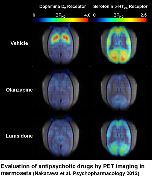
Evaluation of antipsychotic drugs by PET imaging in conscious common marmosets
Patients with schizophrenia suffer from three major symptoms, positive (delusion and hallucination), negative (blunted affects and avolition) symptoms, and cognitive dysfunction. Disorders in dopaminergic and serotonergic neuron are hallmark of schizophrenia. Antagonistic action of many antipsychotic drugs to dopamine D
2 receptor is considered to be essential for the treatment of positive symptom in schizophrenia. Efficacy on other symptoms and less side effects are key features of second-generation antipsychotic drugs, which are thought to be mediated by actions at other receptors, such as serotonin 5-HT
1A , 5-HT
2A , and 5-HT
7 receptors.
PET imaging enables us to confirm target engagement of psychiatric drugs in brain, and is often used to investigate the optimal dose range and mechanism of action of new candidate drugs in the process of in clinical drug development. Our research team, in collaboration with Dainippon Sumitomo Pharma, evaluated the target engagement of antipsychotic drugs by PET imaging using conscious common marmosets, which method we previously established.
In this study, we compared the target engagements of two kinds of antipsychotic drugs, lurasidone, and olanzapine, using specific radiotracers for dopamine D
2 and serotonin 5-HT
2A receptors. We found that both drugs showed similar brain D
2 receptor occupancy in proportion to plasma drug levels, but occupancies at serotonin 5-HT
2A receptor differed between two drugs. Lurasidone is known to have excellent profiles of efficacy and safety, and is approved for the treatment of schizophrenia by FDA in U.S.A, recently. These results allow us to elucidate the mechanism of action of lurasidone, and also give us useful information for understanding the pathophysiology of schizophrenia.
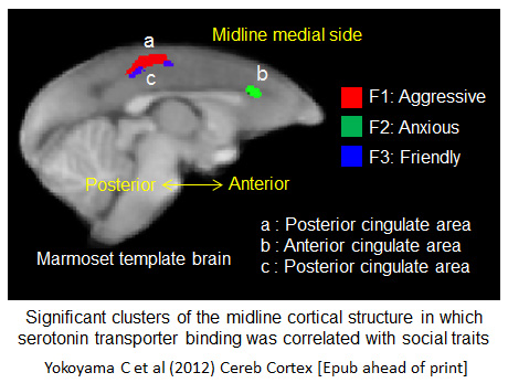
Linkage of serotonin to the social brain underlying 'personality' in monkeys
Evidence indicates that serotonin is involved in regulating emotional behaviors in humans and other primates, but it is unclear which regions of the brain are affected by the serotonergic system in this process. Some evidence suggests that regions of the midline cortex are involved in emotional regulation and social cognition. We have investigated the correlation of behavioural traits in marmosets with serotonin transporter (SERT) binding and neuronal activity in cortical brain regions of the midline cortex.
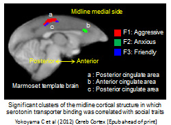
We first classified behavioural traits displayed by male marmosets when they encountered an unfamiliar male, and saw aggressive, anxious and unfriendly traits. We then used positron emission tomography (PET) to assess SERT binding in different brain regions, and saw differential binding that correlated with the behavioural traits. PET also revealed that activity in the same regions, and functional connectivity between them, is altered in different social situations.
The results show that midline cortical regions are involved in serotonergic regulation of social behaviour, and that different subregions play distinct roles in this process, with implications for our understanding of human social cognition and disorders.
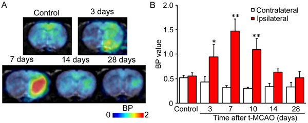
Diagnosis of Disease States of Cerebral Infarction Using Molecular Imaging
Experiments show that 20-Hydroxyeicosatetraenoc acid (20-HETE), presumably playing an autoregulatory function for cerebral blood flow, is transiently and abundantly produced in the rats when they have stroke. The cause of the production of 20-HETE is believed to be a transiently elevated activity of 20-HETE synthetic enzyme in the brain, but the details of the mechanism are still unclear. A clear understanding of the relation between brain disorders and 20-HETE synthase would provide critical information to understand the disease states and prognosis. This time, our research team, in collaboration with Osaka University and Taisho pharmaceutical company, has developed a PET probe [
11C] TROA targeting 20-HETE synthetic enzyme and successfully performed the live imaging of it.
[
11C]TROA is a 20-HETE inhibitor of N'(4-dimethylaminohexyloxy) phenyl imidazole (TROA) labeled by a
11C radioisotope. The PET study using normal rats showed high concentration of [
11C]TROA on kidneys, livers and other organs which are known to have abundant 20-HETE synthetic enzyme under normal conditions. On rat cerebral infarction models, [
11C]TROA was recognized to highly concentrate in the ipsilateral hemisphere of the brain, peaking on day 7 after strokes. This indicates a transient elevation of the activity of 20-HETE synthase following cerebral infarctions.
These results suggest that changes in the activity of 20-HETE synthase are observable by the PET study and such observation will serve as a possible critical diagnostic method to establish the most suitable treatment strategy.
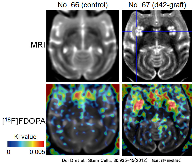
Examination of the Changes in Function of ES Cells Grafted in the Brain Using Live Imaging
Recent advancement in stem cell study such as ES cells and iPS cells has led to the expectation that regenerative therapy with transplantation for Parkinson's disease will be realized to replace lost dopaminergic neurons. However, little is known of the suitable differentiation state which keeps cells safest and produces large treatment effects for Parkinson's disease.

This time, our research team, in collaboration with researcher Daisuke Doi and Associate Professor Jun Takahashi (Center for iPS Cell Research and Application, Kyoto University) compared the growth and function of hESC-derived cells with different stages of neural differentiation implanted in the brains of primate models of Parkinson's disease. After transplanting them, we determined the differences among them of tumorigenicity and the capacity to produce dopamine by live imaging of the grafts, using MRI and PET scan. The result indicated that any tumor formed by undifferentiated graft cells would be detectable, and highly-differentiated graft cells will function as dopamine-producing cells without being transformed into tumors. Our histopathological investigation confirmed the fact as well. Note that only highly-differentiated graft cells effectively improved the symptom of such disease.
The finding of our research demonstrates that the graft treatment using ES cells may lead to the establishment of the protocol for controlling the differentiation of graft cells and for treating the disease effectively and safely, as well as indicating that molecular imaging techniques such as MRI and PET are exceptionally useful evaluation methods to noninvasively verify the graft treatment.
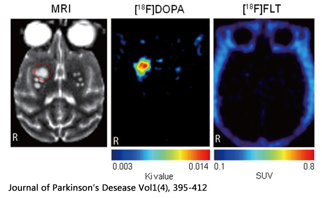
Assessment of efficacy of transplantation therapy with iPSCs by molecular imaging
Parkinson's disease is an intractable disease without a curative treatment because lost nerves cannot be regenerated. However, recent advancement in stem cell study has led to the expectation that regenerative therapy with transplantation will be realized to replace lost dopaminergic neurons.
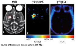
The research team, in collaboration with researcher Tetsuhiro Kikuchi and Associate Professor Jun Takahashi (Center for iPS Cell Research and Application, Kyoto University) explored the culture conditions that allowed human iPSCs to efficiently turn into dopaminergic neurons, then, these neurons were transplanted into a brain of Parkinson's disease model monkey. First of all, the positions of transplanted cells were identified on histological MRI images to examine the post-transplant graft survival. We employed PET imaging in order to determine the properties as functional DA neurons, the results showed an aggressive intake of the dopamine precursor molecules by the transplanted cells, and was confirmed the absence of abnormal proliferation of the transplanted cells. These results demonstrate that the transplanted cells deriving from human iPSCs function as normal neurons with dopamine-producing capacity in the monkey brain. These findings were also verified in histopathological studies.
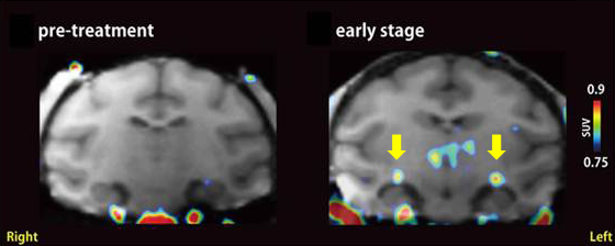
Molecular imaging leads to early detection of glaucoma
Glaucoma is a progressive disease leads to visual deficit, and early detection and therapy of glaucoma are crucial.
Our research team, in collaboration with Professor Hara's team at Gifu Pharmaceutical University, investigated what actually happens in the brains of glaucoma in an experimental glaucoma model monkey using PET.

We focused on finding the inflammatory reaction, particularly, activated microglia, which is known as one of important immune cells in the brain. PET imaging in the glaucomatous monkey with its early-stage revealed that microglia-associated PET probes were accumulated in the deep in the brain, at the lateral geniculate body. This microglial activation was also accompanied by shrinkage and degeneration of neurons in the lateral geniculate body in a histopathological examination. These results suggest the possibility that glaucoma, which has been considered to be a disease of eye, could be correctly diagnosed by brain PET imaging at the initial stage of the disease. Glaucoma is a slowly progressive disease of eye, and is the leading cause of blindness. Early detection of glaucoma is crucial for preventing its progress and for reducing the blind.
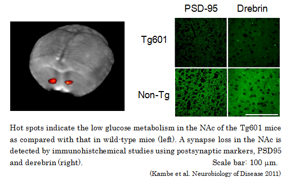
Abnormality of glucose metabolism of nucleus accmubens in tauopathy model mouse detected by PET imaging system under conscious condition
We have found that aged Tg601 mouse, a newer model of tauopathy, showed low glucose metabolism in the nucleus accmubens (NAc) measured by brain PET imaging under conscious condition (Collaboration with Drs. Motoi and Kambe in Juntendo University).
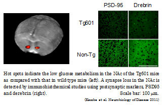
Tau protein has been known to stabilize constitutive axonal microtubules. Tauopathies are neurodegenerative disease such as Alzheimer's disease (AD) and frontotemporal lobar degeneration (FTLD) characterized by the aggregation of hyperphosphorylated tau protein, which leads to the formation of neurofibrillary tangles in neurons and subsequent neuronal death. Although tauopathies are thought to be caused by the mutation of tau gene, some patients did not have any genetic problem of the tau. To clarify whether neuronal accumulated naïve tau protein results in neurodegeneration, we have developed a tauopathy model mouse (Tg601 mouse) by overexpressing a wild-human tau protein (2N4R type) in neuron. The Tg601 mice showed learning deficit and impaired anxiety in the aged period. Using [
18F]FDG-PET imaging method which we have previously established, we found that the aged Tg601 mouse has a low glucose metabolism in the nucleus accmubens (NAc). Interestingly, a synapse loss was also shown in the NAc by immunohistochemical staining. Our in vivo imaging method is really valuable for pathophysiological studies in various genetically manipulated mice.
Successful Live Imaging of COX-1 Enzyme Revealing the Pathogenic Mechanism of Neuroinflammation
COX is the enzyme which produces an inflammation agent and has gained attention as a target for diagnosis and therapy for inflammatory diseases. There are two distinct isoforms (COX-1, COX-2), however, how COXs are specifically related to the incidence of neurodegenerative disease is still unknown.
We labeled a derivative of ketoprofen, which shows a strong blocking effect to COX-1 by
11C, and then develop a new PET probe (
11C-ketoprofen methyl ester;
11C-KTP-Me). Next, the PET probe was shown to specifically recognize COX-1 in the brain by the study of genetically-modified mice lacking COX-1 or COX-2 gene. Furthermore, increase in the accumulation of
11C-KTP-Me in the corresponding inflammation region in the rats with neuroinflammation could be observed by PET imaging. When these rats were observed in detail at the cellular level, increased activation of immune competent cells called microglia, by inflammation reaction together with the expression of COX-1, could be identified. The PET imaging technique for COX-1, as a new bio-marker, can be used to evaluate the degree and progress of neuroinflammation, and is expected to contribute to the identification of the condition, diagnosis, and treatment of neurodegenerative diseases, such as Alzheimer's and Parkinson's diseases.
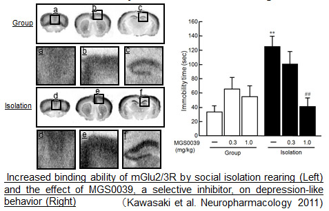
Abnormal neurotransmission in depression discovered
A new study on mGlu2/3R conducted by our research team, in collaboration with Professor Matsuda's team at Osaka University, has found the abnormal mGlu2/3R function in depressive behavior induced by social isolation rearing.

Metabotropic glutamate receptor (mGluR)2/3 is abundantly localized in forebrain regions, in which emotional states are critically controlled. Here we examined the effects of social isolation-rearing on the binding of mGluR2/3 antagonist [3H]LY341495 to mGluR2/3 in the mouse brain. We demonstrated that social isolation rearing increased the binding activity of [3H]LY341495 in the prefrontal cortex and hippocampus of mice. We also found that the mGluR2/3 antagonist MGS0039 reversed the increased immobility time of isolation-reared mice in the forced swim test. These results suggest that the increased binding activity of mGluR2/3 is involved in the depression-like behavior of the socially isolated mice. Further studies in social isolation-reared mice may contribute to clarification of the pathophysiological roles of mGluR2/3 in depression.
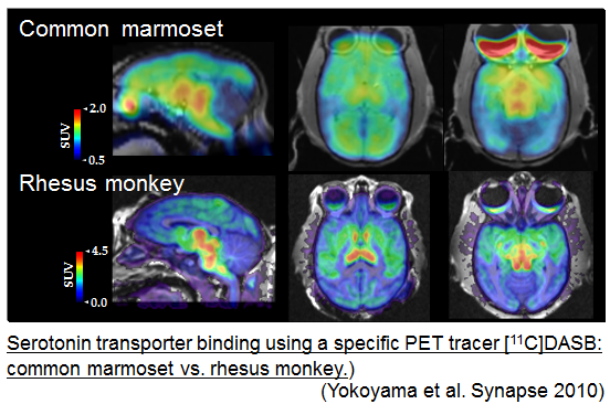
Functional mapping of serotonin transporters in conscious marmoset
We have now established an animal PET imaging method for the conscious common marmoset, which is a small primate used as a biomedical animal model.
PET is useful for investigating in vivo brain function, allowing us to visualize neuronal events associated with the regulatory processes between the genetic and behavioral level. We recruited the common marmoset for PET study as they had been shown to be useful for studying the development of human-like social behavior, such as cooperative sociality. PET with [
11C]DASB, a specific radiotracer for the serotonin transporters, was performed to generate a functional map of the serotonin receptors in the brain of common marmosets under conscious condition. The functional activity and distribution of brain serotonin transporter in conscious common marmosets was vary similar to that of the rhesus monkey and also human.
Establishment of brain imaging method in conscious mouse
We have now established a novel brain PET imaging system for mouse under conscious condition to assess the physiological brain function.
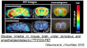
PET with mouse considered to be a significant advantage for studying an intrinsic function of specific molecule under in vivo physiological conditions. However, the animal PET study requires restriction of body movements, therefore is generally conducted under the anesthetized condition. Because anesthetics reduce neural activities and metabolism, an appropriate method of PET imaging under conscious condition is quite important and is practically required for the physiological in vivo brain imaging study. Here, we have established a mouse in vivo imaging system and have found low cerebral glucose metabolic rate (cMRglu) by isoflurane anesthesia is mainly due to decreased hypophosphorylation rate (k3 value), but not glucose transportation rate (K1 value). This method will open avenues for research of in vivo functional brain molecular imaging with the genetically manipulated mice.
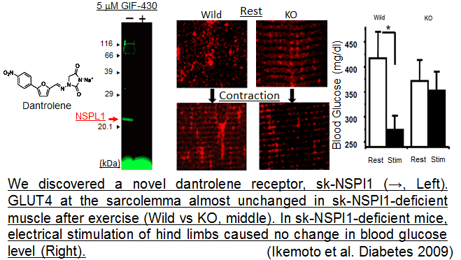
NSPl1; a new factor in the GLUT4 translocation
We have first demonstrated that a skeletal-type neuroendocrine-specificprotein-like 1(sk-NSPl1) in the skeletal muscle cells involves in the GLUT4 translocation and function as a regulator of the blood glucose concentration in a whole body.
Exercise-induced glucose uptake (EIGU) into skeletal muscle cells have been thought to be one of the most important regulatory system of blood glucose level in a body. We have discovered that dantrolene, which inhibits EIGU, can bind to sk-NSPl1. Sk-NSPl1 plays an important role in GLUT4 translocation induced by contraction/exercise. The sk-NSPl1 may also be involved in the regulation of glucose levels in the whole body.
Further study with PET molecular imaging might lead to the understanding of molecular abnormality and the development of preventive or therapeutic agents for various types of lifestyle-related diseases, including type 2 diabetes.
![Search[Google]](../common/image/hmenu_search.png)

![CMIS[RIKEN Center for Molecular Imaging Science]RIKEN Kobe Institute Center for Molecular Imaging Science](../common/image/hlogo_txt.png)